Babesiosis

Babesiosis is a malaria-like parasitic disease caused by infection with Babesia, a genus of protozoal piroplasms. After trypanosomes, Babesia is thought to be the second most common blood parasites of mammals, and they can have a major impact on health of domestic animals in areas without severe winters. Human babesiosis is uncommon, but reported cases have risen recently because of expanded medical awareness.
Symptoms and signs
Most cases of Babesia infection are asymptomatic, but can include mild fevers and diarrhea. The symptoms are often unnoticed or unexplained. In more severe cases, there are symptoms similar to malaria, with fevers up to 40.5°C (105°F), shaking chills, and severe anemia (hemolytic anemia). Organ failure may follow, including adult respiratory distress syndrome. Severe cases occur mostly in people who have had their spleen removed surgically. Severe cases are also more likely to occur in the very young, very old, and persons with immunodeficiency, such as HIV/AIDS patients.
A reported increase in babesiosis diagnoses in the 2000s is thought to be caused by more widespread testing and higher numbers of people with immunodeficiencies coming in contact with ticks, the disease vector. Little is known about the occurrence of Babesia species in malaria-endemic areas, where Babesia can easily be misdiagnosed as Plasmodium.
Pathophysiology
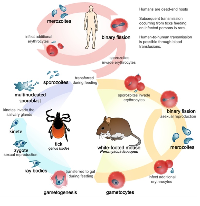
Babesia life cycle
Babesia parasites reproduce in red blood cells, where they can be seen as cross-shaped inclusions (four merozoites asexually budding, but attached together forming a structure looking like a "Maltese cross") and cause hemolytic anemia, quite similar to malaria.
Unlike the Plasmodium parasites that cause malaria, Babesia species lack an exoerythrocytic phase, so the liver is usually not affected.
In animals, Babesia canis rossi, Babesia bigemina, and Babesia bovis cause particularly severe forms of the disease, including a severe haemolytic anaemia, with positive erythrocyte-in-saline-agglutination test indicating an immune-mediated component to the haemolysis. Common sequelae include haemoglobinuria "red-water", disseminated intravascular coaguation and "cerebral babesiosis" caused by sludging of erythrocytes in cerebral capillaries.
In bovine species, the organism causes hemolytic anemia, so an infected animal will show pale mucous membranes initially. As the levels of billirubin (a byproduct of red blood cell lysis) continue to increase, the visible mucous membranes will become yellow in color (icterus) due to the failure of the liver to metabolise the excess bilirubin. Hemoglobinuria will be seen due to excretion of red-blood-cell lysis byproducts via the kidneys. Fever of 40.5°C (105°F) develops due to release of inflammatory byproducts.
Diagnosis
A high index of suspicion is necessary to diagnose babesiosis. It develops only in patients who live in or travel to an endemic area or receive a contaminated blood transfusion within the preceding 9 weeks, so this aspect of the medical history is vital. Babesiosis may be suspected when a person with such an exposure history develops persistent fevers and hemolytic anemia. The definitive diagnostic test is the identification of parasites on a Giemsa-stained thin blood smear.
So-called "Maltese cross formations" on the blood film are essentially diagnostic of babesiosis, since they are not seen in malaria, the primary differential diagnosis. Careful examination of multiple smears may be necessary, since Babesia may infect less than 1% of circulating red blood cells and thus be easily overlooked.
Serologic testing for antibodies against Babesia (both IgG and IgM) can detect low-level infection in cases with a high clinical suspicion, but negative blood film examinations. Serology is also useful for differentiating babesiosis from malaria in cases where people are at risk for both infections. Since detectable antibody responses require about a week after infection to develop, serologic testing may be falsely negative early in the disease course.
A polymerase chain reaction (PCR) test has been developed for the detection of Babesia from the peripheral blood. PCR may be at least as sensitive and specific as blood film examination in diagnosing babesiosis, though it is also significantly more expensive. Most often, PCR testing is used in conjunction with blood film examination and possibly serologic testing.
Other laboratory findings include decreased numbers of red blood cells and platelets on complete blood count.
In animals, babesiosis is suspected by observation of clinical signs (haemoglobinuria and anaemia) in animals in endemic areas. Diagnosis is confirmed by observation of merozoites on thin film blood smear examined at maximum magnification under oil using Romonovski stains (methylene blue and eosin). This is a routine part of the veterinary examination of dogs and ruminants in regions where babesiosis is endemic.
Babesia canis and B. bigemina are "large babesias" that form paired merozoites in the erythrocytes, commonly described as resembling "two pears hanging together", rather than the "Maltese Cross" of the "small babesias". Their merozoites are approximately twice the size of small babesias.
Cerebral babesiosis is suspected in vivo when neurological signs (often severe) are seen in cattle that are positive for B. bovis on blood smear, but this has yet to be proven scientifically. Outspoken red discolouration of the grey matter post mortem further strengthens suspicion of cerebral babesiosis. Diagnosis is confirmed post mortem by observation of babesia-infected erythrocytes sludged in the cerebral cortical capillaries in a brain smear.
Treatment
Most cases of babesiosis resolve without any specific treatment. For ill patients, treatment is usually a two-drug regimen, quinine and clindamycin. As these drugs are often poorly tolerated, recent evidence suggests a regimen of atovaquone and azithromycin can be equally effective. In life-threatening cases, exchange transfusion is performed. In this procedure, the infected red blood cells are removed and replaced with uninfected ones.
Epidemiology
Babesiosis is a vector-borne illness usually transmitted by Ixodes scapularis ticks. Babesia microti uses the same tick vector as Lyme disease and ehrlichiosis, and may occur in conjunction with these other diseases. The organism can also be transmitted by blood transfusion. Ticks of domestic animals, especially Rhipicephalus (Boophilus) microplus and R. (B.) decoloratus transmit several species of Babesia to livestock animals, causing considerable economic losses to farmers in tropical and subtropical regions.
In North America, the disease is primarily found in eastern Long Island, Fire Island, Nantucket Island and Martha's Vineyard off of the coast of Massachusetts. More generally, it can be found in the northern midwestern and New England states. It is sometimes called "The Malaria of The Northeast." Cases of babesiosis have been reported in a wide range of European countries. Disease in Europe is usually due to infection with Babesia divergens, while in the United States Babesia microti andBabesia duncani are the species most commonly associated with human disease. Babesiosis has also been observed in Korea.
Babesiosis has emerged in Lower Hudson Valley, New York since 2001.
In Australia, babesiosis has recently been proven an endemic infection with evidence of both B. duncani and B. microti along the eastern coastline of the continent as a tick-borne infection. A similar disease in cattle, commonly known as tick fever, is spread by Babesia bovis and B. bigemina in the introduced cattle tick Rhipicephalus microplus. This disease is found in eastern and northern Australia.
History
The disease is named for the genus of the causative organism, which was named after the Romanian bacteriologist Victor Babeş.
Equinebabesiosis is also known as piroplasmosis (from the Latin piro, meaningpear + German plasma, a thing formed).
In other animals
Veterinary treatment of babesiosis does not normally use antibiotics. In animals, diminazen (Berenil), imidocarb or trypan blue would be the drugs of choice for treatment of B. canis rossi (dogs in Africa), B. bovis, and B. bigemina (cattle in Southern Africa).
A vaccine is effective against B. canis canis (dogs in the Mediterranean region), but is ineffective against B. c. rossi. Babesia imitans causes a mild form of the disease that frequently resolves without treatment (dogs in southeast Asia).
