Canine distemper
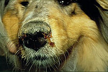
Dog infected with canine distemper
Group: Group V ((-)ssRNA)
Order: Mononegavirales
Family: Paramyxoviridae
Genus: Morbillivirus
Species: Canine distemper virus
Canine distemper is a viral disease that affects animals in the families Canidae, Mustelidae, Mephitidae, Hyaenidae, Ailuridae, Procyonidae, Pinnipedia, some Viverridae and Felidae (though not domestic cats; feline distemper or panleukopenia is a different virus exclusive to cats). It is most commonly associated with domestic animals such as dogs and ferrets, although it can infect wild animals as well. It is a single-stranded RNA virus of the family paramyxovirus, and thus a close relative of measles and rinderpest. Despite extensive vaccination in many regions, it remains a major disease of dogs.
Etymology
The origin of the word distemper is from the Middle English distemperen, meaning to upset the balance of the humors, which is from the Old French destemprer, meaning to disturb, which is from the Vulgar Latin distemperare: Latin dis- and Latin temperare, meaning to not mix properly.
History
Although very similar to the measles virus, canine distemper virus (CDV) seems to have appeared more recently, with the first case described in 1905 by French veterinarian Henri Carré.It was first thought to be related to the plague and typhus, and was attributed to several species of bacteria. It now affects all populations of domestic dog and some populations of wildlife. The first vaccine against canine distemper was developed in 1923 and 1924 by an Italian named Puntoni, although he did not use a large population of dogs for his trials, his work shows dogs can be vaccinated against this disease producing solid immunity. Commercial vaccine was developed in 1950, yet due to limited use, the virus remains prevalent in many populations. The domestic dog has largely been responsible for introducing canine distemper to previously unexposed wildlife, and now causes a serious conservation threat to many species of carnivores and some species of marsupials. The virus contributed to the near-extinction of the black-footed ferret. It also may have played a considerable role in the extinction of the thylacine (Tasmanian tiger) and recurrently causes mortality among African wild dogs. In 1991, the lion population in Serengeti, Tanzania, experienced a 20% decline as a result of the disease. The disease has also mutated to form phocid distemper virus, which affects seals.
Infection
The virus, a single-stranded negative RNA, can cause systemic infection in the host carnivore. Puppies from three to six months old are particularly susceptible. CDV spreads through aerosol droplets and through contact with infected bodily fluids, including nasal and ocular secretions, feces, and urine, six to 22 days after exposure. It can also be spread by food and water contaminated with these fluids. The time between infection and disease is 14 to 18 days, although a fever can appear from three to six days after infection.
Canine distemper virus tends to orient its infection towards the lymphoid, epithelial, and nervous tissues. The virus initially replicates in the lymphatic tissue of the respiratory tract. The virus then enters the blood stream and infects the respiratory, gastrointestinal, urogenital epithelial, and central nervous systems, and optic nerves. Therefore, the typical pathologic features of canine distemper include lymphoid depletion (causing immunosuppression and leading to secondary infections), interstitial pneumonia, encephalitis with demyelination, and hyperkeratosis of the nose and foot pads.
The mortality rate of the virus largely depends on the immune status of the infected dogs. Puppies experience the highest mortality rate, where complications such as pneumonia and encephalitis are more common. In older dogs that develop distemper encephalomyelitis, vestibular disease may present. Around 15% of canine inflammatory central nervous system diseases are a result of CDV.
Disease progression
The virus first appears in bronchial lymph nodes and tonsils two days after exposure. The virus then enters the blood stream on the second or third day. A first round of acute fever tends to begin around three to eight days after infection, which is often accompanied by a low white blood cell count, especially of lymphocytes, as well as low platelet count. These signs may or may not be accompanied by anorexia, a runny nose, and discharge from the eye. This first round of fever typically recedes rapidly within 96 hours, and then a second round of fever begins around the 11th or 12th day and lasts at least a week. Gastrointestinal and respiratory problems tend to follow, which may become complicated with secondary bacterial infections. Inflammation of the brain and spinal cord, otherwise known as encephalomyelitis, is either associated with this, subsequently follows, or comes completely independent of these problems. A thickening of the footpads sometimes develops, and vesicularpustular lesions on the abdomen usually develop. Neurological symptoms typically are found in the animals with thickened footpads from the virus. About half of sufferers experience meningoencephalitis.
Gastrointestinal and respiratory symptoms
Commonly observed signs are a runny nose, vomiting and diarrhea, dehydration, excessive salivation, coughing and/or labored breathing, loss of appetite, and weight loss. When and if the neurological symptoms develop, incontinence may ensue.
Neurological symptoms
The symptoms within the central nervous system include a localized involuntary twitching of muscles or groups of muscles, seizures often distinguished by salivation, and jaw movements commonly described as "chewing gum fits", or more appropriately as "distemper myoclonus". As the condition progresses, the seizures worsen and advance to grand mal convulsions, followed by death of the animal. The animal may also show signs of sensitivity to light, incoordination, circling, increased sensitivity to sensory stimuli such as pain or touch, and deterioration of motor capabilities. Less commonly, it may lead to blindness and paralysis. The length of the systemic disease may be as short as 10 days, or the start of neurological symptoms may not come until several weeks or months later. Those few that survive usually have a small tic or twitch of varying levels of severity. With time, this tic will usually diminish somewhat in its severity.
Lasting symptoms
A dog that survives distemper will continue to have both nonlife-threatening and life-threatening symptoms throughout their lifespans. The most prevalent nonlife-threatening symptom is hard pad disease. This is when a dog experiences the thickening of the skin on the pads of its paws, as well as the end of its nose. Another lasting symptom commonly is enamel hypoplasia. Puppies, especially, will have damage to the enamel of teeth that are not completely formed or those that have not yet grown through the gums. This is a result of the virus killing the cells responsible for manufacturing the tooth enamel. These affected teeth tend to erode quickly.
Life-threatening symptoms usually include those due to the degeneration of the nervous system. Dogs that have been infected with distemper tend to suffer a progressive deterioration of mental abilities and motor skills. With time, the dog can acquire more severe seizures, paralysis, reduction in sight and incoordination. These dogs are usually humanely euthanized, due to the immense pain and suffering they face.
Diagnosis
The above symptoms, especially fever, respiratory signs, neurological signs, and thickened footpads found in unvaccinated dogs strongly indicate canine distemper. However, several febrile diseases match many of the symptoms of the disease and only recently has distinguishing between canine hepatitis, herpes virus, parainfluenza and leptospirosis been possible. Thus, finding the virus by various methods in the dog's conjunctival cells gives a definitive diagnosis. In older dogs that develop distemper encephalomyelitis, diagnosis may be more difficult, since many of these dogs have an adequate vaccination history.
An additional test to confirm distemper is a brush border slide of the bladder transitional epithelium of the inside lining from the bladder, stained with Dif-Quick. These infected cells have inclusions which stain a carmine red color, found in the paranuclear cytoplasm readability. About 90% of the bladder cells will be positive for inclusions in the early stages of distemper.
Prevention
A number of vaccines against canine distemper exist for dogs (ATCvet code: QI07AD05 and combinations) and domestic ferrets (QI20DD01), which in many jurisdictions are mandatory for pets. The type of vaccine should be approved for the type of animal being inoculated, or else the animal could actually contract the disease from the vaccine. Infected animals should be quarantined from other dogs for several months due to the length of time the animal may shed the virus.The virus is destroyed in the environment by routine cleaning with disinfectants, detergents, or drying. It does not survive in the environment for more than a few hours at room temperature (20–25°C), but can survive for a few weeks in shady environments at temperatures slightly above freezing. It, along with other labile viruses, can also persist longer in serum and tissue debris.
Treatment
Until recently, canine distemper has been associated with a long history of pessimism with respect to treatment of infected animals and the disease was usually assumed to have a poor prognosis. Most care offered was only palliative, geared toward easing the suffering. Several factors had an important role in maintaining the status quo.
Research and funding, for the most part, have focused on vaccination rather than on finding a cure for distemper.
In an outdated theory, the injuries that occurred were the result of a strictly autoimmune reaction, with the thought being that initially the canine distemper virus was introduced, but then subsequently eliminated, and that the cytokines continued to attack and damage healthy tissue in the absence of a current pathogen. Based on that theory, anti-inflammatory and immunosuppressive drugs have been prescribed by some veterinarians in an attempt to bring the effects of the condition under control, but this did not succeed.
It was later considered that the action of macrophages on infected nerve cells indicated the autoimmune reaction was likely a direct consequence of the presence of the virus. Often, owners seek expert help only when the disease is in its advanced stages (nervous phase) due to the nonspecific earlier signs and prescription of anti-inflammatory drugs (which are usually corticosteroids) undermine the immune system of the animal, allow the proliferation of the virus, and the autoimmune reaction increases as a means of containment of infected cells.
The first references to suggest effective treatments for similar viruses could be effective for canine distemper arose when studies found canine distemper was a disease comparable to measles, and infected animals could be used to develop new technologies for treatment of measles. The question of whether the reciprocal would be true was resolved when studies assessed the efficacy of traditional treatments for measles, some of which were successfully applied to animals with distemper.
Vitamin A and ribavirin as a treatment of distemper (and measles) are under evaluation and the modes of action remain unexplained. Some experiments suggest vitamin A and ribavirin could be effective as a treatment, but to date is not commonly employed in practice. Some evidence points to an indirect action, such as confirming a reduction in the amounts of vitamin A during infection, pointing to the hypothesis that it is raw material for some mechanism of resistance to infection. That the anti-infective characteristic is not specific to vitamin A is a mystery; however, there was no doubt about its effectiveness in this particular experiment, action mechanisms elucidated or not.
The adoption of ribavirin as a possible treatment for canine distemper followed the same steps as vitamin A; it was the principle used in cases of subacute sclerosing panencephalitis under measles. The first verification of the effectiveness occurred in vitro. The distemper virus was observed to be very susceptible to ribavirin, and 0.02 to 0.05 micromols are needed to induce its mechanism of error catastrophe and the inhibitory effect on virus replication by 50%.
The main concern in the use of ribavirin was the result of its interaction with the blood–brain barrier. As the brain is an immunologically privileged area, the concern was the capacity of ribavirin to overcome this barrier. In a study using mice with encephalitis due to measles, once the virus has become established in the nervous phase, the blood–brain barrier fails, in a way, reducing the restriction to the action of the ribavirin in these areas. The verification of all these results in vivo resulted in an effectiveness of 80% in animals that had already reached the nervous phase of viral infection. The application of ribavirin demands a close monitoring of the animal due the risk of leukopenia; also, the ingestion of long-chain tryglicerides (fats) are needed to better absorb the drug, and for preservation of gastric tissues, which are quite susceptible to it.
The medical and veterinary communities widely accept, while possible treatments are still being explored into their efficacy, prevention using vaccination is the most reliable way of preventing disease spread among the population.
Relation to Paget's disease
Paramyxoviruses, such as CDV, measles, respiratory syncytial virus, simian virus 5, and parainfluenza virus type 3, have long been suspected as the causative agents of Paget's disease, a focal destructive disease of bone. Most studies, however, have pointed more directly at CDV and measles. A virus detection technique, in situ RT-PCR, has found CDV in 100% of Paget's disease samples, whereas other virus detection techniques have been less accurate.
Prevalence
The prevalence of canine distemper in the community has decreased dramatically due to the availability of vaccinations. However, the disease continues to spread among unvaccinated populations, such as animal shelters and pet stores. This provides a great threat to both the rural and urban communities throughout the United States, affecting both shelter and domestic canines. Despite the effectiveness of the vaccination, outbreaks of this disease continue to occur nationally. In April of 2011, the Arizona Humane Society released a valley-wide pet health alert throughout the Phoenix, Arizona area.
Contributing factors
Outbreaks of canine distemper continue to occur throughout the United States and elsewhere, and are caused by many factors. These factors include the overpopulation of dogs and the irresponsibility of pet owners. The overpopulation of dogs is a national problem that organizations such as the Humane Society and ASPCA face every day. This problem is even greater within areas such as Arizona, due to the vast amount of rural land. An unaccountable number of strays that lack vaccinations reside in these areas, and are therefore more susceptible to diseases such as canine distemper. These strays act as a host for the virus, spreading it throughout the surrounding area including urban areas. Puppies and dogs that have not received their shots can then be infected if in a place where many dogs interact, like a dog park.
The irresponsibility of pet owners is another factor that contributes to ongoing outbreaks of canine distemper. Puppies begin vaccination beginning at six to eight weeks of age, and then continue getting the “booster shot” every two to four weeks until they are 16 weeks of age. Without the full series of shots, the vaccination will not provide prevention against the virus. Dog owners often vaccinate their dogs with the first, but do not finish the series. These dogs are not protected against the virus, so are susceptible to canine distemper infection, continuing the downward spiral that leads to outbreaks throughout the country.
